Mental Illness And Diagnostic Brain Imaging Systems:
CT, MRI, SPECT, PET, EEG, QEEG, fMRI
Neuroscientists and psychiatrists admit they cannot find the cause of mental illness with brain-imaging techniques.
|
|
Brain imaging systems are very crude devices in that they merely measure blood flow in the brain which is assumed to correspond with electrical activity of the neurons in the brain. They cannot measure what is being thought or if there is a problem with thinking patterns. They are of no use in determining the cause of mental illness. Mental illnesses are rooted in the spirit, not the physical brain. The brain is a mere interface between the spirit and the body. The body is dead without the spirit. The spirit will continue to exist and think after the death of the body. |
Introduction:
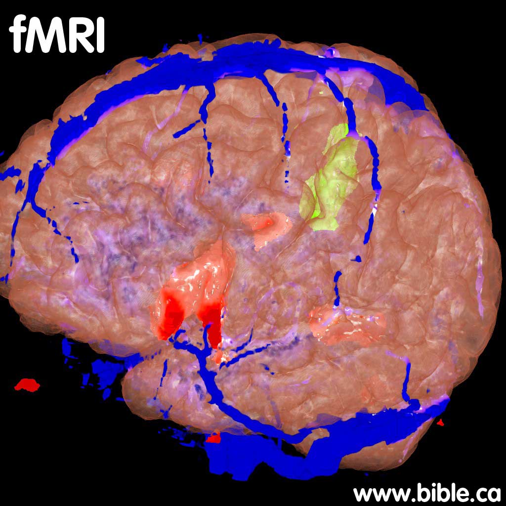
1. Brain imaging systems are assumed to measure total electrical current inside the brain by measuring blood flow. They can also pinpoint where the current is flowing in the different parts of the brain. What and fMRI cannot do, for example, is tell you anything about what is the person is actually thinking. Brain imagery is based upon the theoretical relationship between neuronal activity and regional blood flow. What is measured is the amount of blood flow in parts of the brain. The theory then translates the amount of blood flow into electrical activity, which is then used to determine where we do our thinking. The theory: Blood flow increases in areas of the brain where neuronal activity increases. But an fMRI does not directly measure electrical activity in different parts of the brain, it simply measures blood flow.
a. This is what EEG (Electroencephalography), QEEG (Quantitative Electroencephalography), PET (positron emission tomography), MRI (magnetic resonance imaging) and fMRI (functional magnetic resonance imaging) machines do. They measure electrical activity but cannot interpret thought and emotion.
b. An ammeter: Ammeter is used by electricians to tell the flow of electricity (current) inside a single wire or a group of wires. For example, ammeters can be used to measure the flow of electrical current inside a trunk phone wire that can service 10,000 different phone lines. Now the ammeter can easily measure the total current, even isolate which phone lines are being used and which are not, but it cannot tell you what is being communicated!
c. A light meter: A light meter can measure the amount of electricity in a fibre optics wire, but it cannot interpret the intelligence being communicated.
d. A disk drive light: On most computers and laptops, where is a little light that goes on when the disk drive is reading or writing information. When it is on, you can tell something is happening, but you cannot tell WHAT is happening.
- Technically, an fMRI measures the change in magnetization between oxygen-rich and oxygen-poor blood and then assumes a correlation to electrical activity. It is a rather crude device and of no value in determining thinking patterns or even if thought has its origin in one part of the brain.
- The general public are mislead to believe by mental health organizations that mental illness can be seen in brain scans. The truth is that Neuroscientists and psychiatrists admit they cannot find the cause of mental illness with brain-imaging techniques, but they expect some day to find the proof!
- Brain imaging systems can distinguish between normal and abnormal brain circuitry as in epilepsy. But this is a physical problem with wiring, not a spiritual problem of the mind. This sharply contrasts with the fact that there are no differences in the brains of schizophrenics, except those changes from psychiatric drug induced chemical imbalances.
- "Epilepsy: The most important use of EEG continues to be in the diagnosis of seizure disorders. No other brain abnormality has an electrophysiologic pattern as distinctive as epilepsy (Duffy 1988). Epilepsy is found in approximately 0.3"k-0.6% of adults in the general population (Anderson et al. 1999). The presence of spikes (defined as a potential with a duration less than 70 msec), sharp waves (duration of 70-200 msec) and polyspikes, frequently followed by a slow wave, are often seen interictally in epileptic patients (Aminoff 1986; Goodin and Aminoff 1984)." (Textbook of Neuropsychiatry and Clinical Neurosciences, Yudofsky, Hales, 2002 AD, p 205)
- Biological Psychiatrists view man as nothing more than a pile of chemicals and that the cause of mental illness is a broken brain. They believe that the key to understanding mental illness is brain imaging. They are wrong.
- This is what EEG (Electroencephalography), QEEG (Quantitative Electroencephalography), PET (positron emission tomography), MRI (magnetic resonance imaging) and fMRI (functional magnetic resonance imaging) machines do. They measure electrical activity but cannot interpret thought and emotion.
- Stated simply: The error of Biological Psychiatrists is that you can cannot determine the cause of mental illness by using fMRI. This is like a Macintosh computer user x-raying the Mac CPU to determine why the Mac software keeps crashing.
- While science fiction often shows machines that can "read the thoughts of the mind". Such is impossible and no technology is even remotely capable of mind reading.
A. Understanding Brain imaging systems:
|
|
|
|
|
Brain imaging systems Mental Illness and Brain imaging systems: CT, MRI, SPECT, PET, EEG, QEEG, fMRI Brain imaging systems are very crude devices in that they merely measure electrical activity of the neurons in the brain. They cannot measure what is being thought or if there is a problem with thinking patterns. |
|
|
|
|
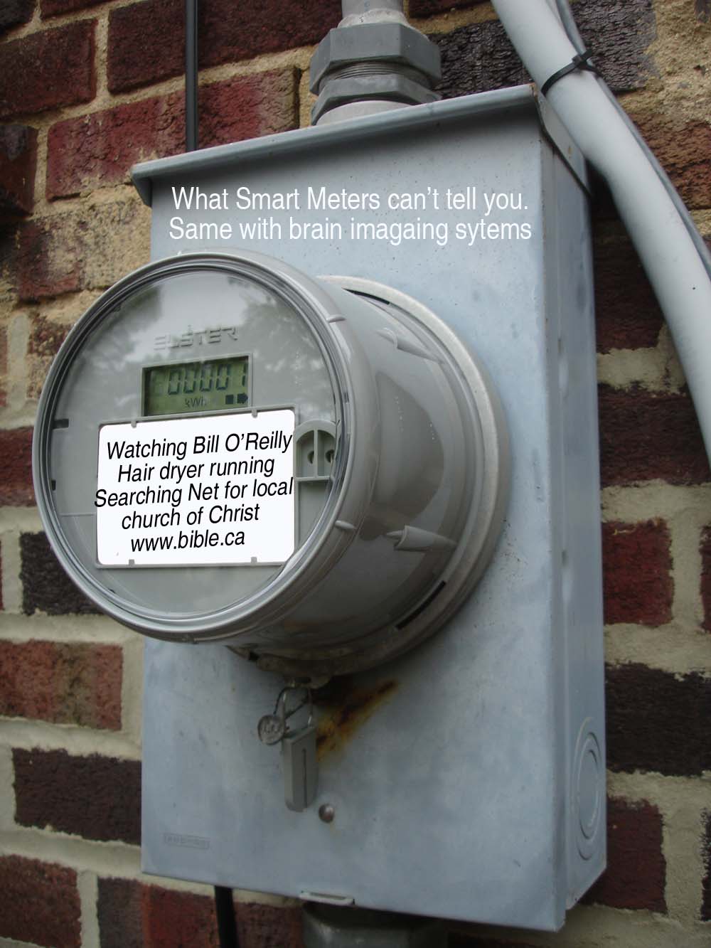
Brain imaging systems merely measure electrical current like a "smart meter" but they cannot read thoughts. A household smart meter cannot tell you what the electricity is doing inside. They tell you how much power, not what the power is doing! Watching Bill O'Reilly |
|
|
|
|
|
|
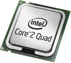
Trying to understand the mind by examining the brain, is like pulling the CPU chip (main brain) out of a computer and looking at the transistors, resisters and diodes to understand Windows XP. The CPU could be running a wide variety of programs, just like the brain can think a wide range of thoughts. If the computer crashes, it is never the CPU's fault, but a bug in the software. Likewise a "nervous breakdown" is not caused by chemical imbalances in the brain, but the spirit. A disk drive light: On most computers and laptops, where is a little light that goes on when the disk drive is reading or writing information. When it is on, you can tell something is happening, but you cannot tell WHAT is happening. "The light is on, but no idea what is happening in the home!" |
|
|
|
|
Ammeter is used by electricians to tell the flow of electricity (current) inside a single wire or a group of wires. For example, ammeters can be used to measure the flow of electrical current inside a trunk phone wire that can service 10,000 different phone lines. Now the ammeter can easily measure the total current, even isolate which phone lines are being used and which are not, but it cannot tell you what is being communicated! Brain imaging systems are like a passive inductive ammeter that merely measures electricity between neurons, not the thoughts. |
|
|
|
|

B. No proof of mental illness in the brain:
|
|
Neuroscientists and psychiatrists admit they cannot find the cause of mental illness with brain-imaging techniques. Take special note of phrases like: "not as yet been able", "impressive amount of experimental data", "have the potential" "possibility of symptom localization in schizophrenia", may eventually provide the basis" |

- "These exciting investigational achievements through laboratory and brain-imaging research, however, have not as yet been able to provide an innovative new basis for the comprehensive diagnostic categorization of classic psychiatric disorders such as schizophrenia and unipolar depression. Indeed, we have not yet even achieved incremental validity. In other words, there is as yet no definitive evidence that any psychiatric laboratory test or brain-imaging mea-sure can provide a comprehensive and clearly incremental improvement to the existent approach to the clinical diagnosis of classic psychiatric illnesses (Morihisa 1991)." (Textbook of Clinical Psychiatry, Hales, Yudofsky, 2003 AD, p 250)
- "Functional brain imaging refers to a class of techniques that non-invasively measure correlates of neural activity. Positron emission tomography (PET) and functional magnetic resonance imaging (fMRI) are the two technologies most commonly used today to study the human brain "in action." The explosion of information about human brain function occurring in the last decade has resulted in large part from these two techniques. In particular, fMRI has gained rapid acceptance because of the widespread availability of MRI scanners and the lack of radioactive exposure. The advent of neuroimaging techniques for probing in vivo human brain function undoubtedly represents a major milestone in the scientific endeavor of understanding the relationship between mental disorders and the brain. The development of the specific tools employed in brain mapping, although fairly recent, has already produced an impressive amount of experimental data, whose potential informational content is most likely being underexploited at the present time (Van Horn and Gazzaniga 2002)." (Textbook of Psychopharmacology, Schatzberg, Nemeroff, 2002 AD, p 163)
- "As for psychiatry, it ought to be clear that, except for the diagnoses of neurological diseases (which are treated by neurologists), no psychiatric diagnosis is, or can be, pathology-driven; instead, all such diagnoses are driven by nonmedical—that is, economic, personal, legal, political, and social—considerations or incentives. Accordingly, psychiatric diagnoses point neither to patho-anatomic or patho-physiological lesions, nor to disease-causative agents, but to human behaviors and human problems, and to the fallible attempts of fallible moral agents to cope with problematic human behaviors." (The Medicalization Of Everyday Life, Thomas Szasz, 2007 AD, p 36)
- "Brain Imaging: Whereas the proposed neuroendocrine tests largely provide an indirect measure of brain activity (e.g., through central effects on endocrine function), brain-imaging techniques have the potential for providing a more direct window on the functioning of the living human brain." (Textbook of Clinical Psychiatry, Hales, Yudofsky, 2003 AD, p237)
- In summary, despite the fact that the physiological and biochemical processes linking the neural activity and the hemodynamic response have not been clarified yet, the empirical relationship between these parameters appears both reliable and reproducible in a variety of con-texts." (Textbook of Psychopharmacology, Schatzberg, Nemeroff, 2002 AD, p 165)
- "fMRI is highly sensitive to picking up changes in activity; however, there is a large amount of intersubject variation, and the specific alterations associated with psychiatric illness are an evolving field. Multimodal imaging through the combination of fMRI, PET, and electromagnetic measurements (electroencephalography, magnetoencephalography) offers the promise of identifying both neuronal and chemical changes related to brain function." (Textbook of Psychopharmacology, Schatzberg, Nemeroff, 2002 AD, p 171)
- "The development of brain-imaging techniques such as CT, MRI, SPECT, and PET have enhanced our understanding of schizophrenia. This technology is allowing us to explore the nature and pattern of brain deficits and examine the possibility of symptom localization in schizophrenia." (Textbook of Clinical Psychiatry, Hales, Yudofsky, 2003 AD, p 426)
- "Nevertheless, the evolving alliance between brain imaging, molecular genetics, and the cognitive (Carter 2001) and basic neurosciences raises the possibility of identifying dysfunctional neural networks in psychiatric illnesses that may eventually provide the basis for enhanced diagnostic, prognostic, and treatment approaches in diseases such as schizophrenia and depression (Morihisa 2001)." (Textbook of Clinical Psychiatry, Hales, Yudofsky, 2003 AD, p 251)
- "Compared with the large number and rapid pace of fMRI studies in psychiatrically healthy subjects, the use of brain imaging in psychiatric illness is more restrained."(Textbook of Psychopharmacology, Schatzberg, Nemeroff, 2002 AD, p 170)
- "Even if it is known [based upon accepted theory which is wrong] that there are major alterations in brain function (e.g., such as in schizophrenia), pinning them down with brain imaging is not easy. " (Textbook of Psychopharmacology, Schatzberg, Nemeroff, 2002 AD, p 170)
C. Thinking and emotions activate almost the entire brain:
- Modern Psychiatry is looking for specific areas of the brain that control specific emotions. This is neo-phrenology and it is wrong. Emotions have their origin in the human spirit. The brain is merely the physical interface between the spirit and the body. All thoughts and emotions trigger large areas of the brain in many different areas.
- "Our findings demonstrate that there is no single "God spot" in the brain located in the temporal lobes. Rather our objective and subjective data suggest that RSMEs [religious/spiritual/mystical experiences] are complex and multidimensional and mediated by a number of brain regions normally implicated in perception, cognition, emotion, body representation, and self-consciousness." (The Spiritual Brain, Mario Beauregard Ph.D., Neuroscientist, 2007, p272)
- "We learned two valuable things from our studies. The results of the two studies, taken together (QEEG and fMRI), dispose of the notion that there is a God spot in the temporal lobes of the brain that can somehow "explain" RSMEs [religious/spiritual/mystical experiences]. The results of our fMRI and QEEG studies suggest that RSMEs are neurally instantiated by different brain regions involved in a variety of functions, such as self-consciousness, emotion, body representation, visual and motor imagery, and spiritual perception. This conclusion correlates well with subjects' descriptions of RSMEs as complex and multidimensional." (The Spiritual Brain, Mario Beauregard Ph.D., Neuroscientist, 2007, p274)
- "The main goal of this functional magnetic resonance imaging (fMRI) study was to identify the neural correlates of a mystical experience. The brain activity of Carmelite nuns was measured while they were subjectively in a state of union with God. This state was associated with significant loci of activation in the right medial orbitofrontal cortex, right middle temporal cortex, right inferior and superior parietal lobules, right caudate, left medial prefrontal cortex, left anterior cingulated cortex, left inferior parietal lobule, left insula, left caudate, and left brain stem. Other loci of activation were seen in the extra-striate visual cortex. These results suggest that mystical experiences are mediated by several brain regions and systems." (Mario Beauregard and V. Paquette, "Neural Correlates of a Mystical Experience in Carmelite Nuns," Neuroscience Letters 405 (2006): 186-90)
D. Understanding brain-imaging techniques:
- "Functional Magnetic Resonance Imaging: Correlates of Neural Activity: It has been known for over 100 years that blood flow to the brain increases in a regionally specific manner, according to mental activity. The father of modern psychology, William James, was aware of observations relating regional brain pulsation to mental activity (James 1890). Paul Broca, known primarily for his observations on the effects of left frontal lesions on language, performed several experiments relating regional brain temperature to cognitive function (Raichle 1998). It was not until the 1950s, however, when Seymour Kety and Louis Sokoloff developed the autoradiographic technique for quantitatively measuring regional blood flow, that specific cognitive functions could be directly mapped in the living brain (Kety 1965). Both PET and fMRI rely on the empirical relationship between neuronal activity and regional blood flow. Stated simply, blood flow increases in areas where neuronal activity increases, and most cognitive neuroscience studies implicitly assume the validity of this relationship. It is easy to see the link in terms of an increased metabolic demand. The activation of a neural circuit is a complex net-work of electrochemical processes that requires energy. The most demanding processes, in terms of energetic expenditure, are related to synaptic (rather than spiking) activity, including the mechanisms of exocytosis, the re-uptake of neurotransmitters, and the restoration of ionic concentrations. The principal energy currency in the brain, as well as in the entire organism, is the adenosine triphosphate (ATP) molecule, whose characteristic phosphate bonds allow the storage and release of energy in a highly efficient manner. To replace the ATP degraded by the increased metabolic demand, a novel contribution of glucose and oxygen is necessary, which is mediated, in ways still largely unknown, by a vascular response that in-creases the delivery of arterial blood to the activated region. This vascular or hemodynamic response to neural activity (i.e., a variation of regional cerebral blood flow [rCBF]) is the quantity that is actually measured in the majority of brain activation studies with both fMRI and PET/SPECT (Arthurs and Boniface 2002; Jueptner and Weiller 1995), represents an indirect assay of neural activity (Villringer and Dirnagl 1995). It is important to note that the hemodynamic response lags behind the actual neural activity by a few seconds. It is also blurred in the spatial (as well as in the temporal) domain, compared with the underlying neural activity, imposing fundamental limits on the spatiotemporal resolution of blood flow methods. A special caveat should be mentioned concerning the interpretation of rCBF results. The measurement of task- related variations of rCBF, although reflecting a change in population synaptic activity, does not provide any clear indication about the sign of the latter, whether excitatory or inhibitory; however, hypotheses have been proposed for and argued against a bias favoring excitatory contributions (Heeger et al. 1999; Tagamets and Horwitz 2001; Waldvogel et al. 2000). The construction of specific inferences about the actual state of activity (actively ex-cited or actively inhibited) of brain regions, characterized by an increase in rCBF during an experimental task, necessitates the integration of information from different sources (electrophysiology, neurochemistry, cytoarchitectonics, etc.). In summary, despite the fact that the physiological and biochemical processes linking the neural activity and the hemodynamic response have not been clarified yet, the empirical relationship between these parameters appears both reliable and reproducible in a variety of con-texts. Simultaneous recordings of neuronal spiking, field potentials, and fMRI suggest that the mean field potential, which represents a weighted average of the input signals of a local neural population, is linearly related to the signal change measured with fMRI (Logothetis et al. 2001)." (Textbook of Psychopharmacology, Schatzberg, Nemeroff, 2002 AD, p 165)
- "Role of Functional Brain Imaging: Functional brain imaging in psychiatry is used primarily as a research tool to elucidate both normal and abnormal brain circuitry. Because of the wide-ranging psychiatric pathologies in both cognitive and affective function, nearly every cognitive process is potentially a target for these techniques. Memory, mood, attention, language, and motor function constitute some of the large domains of cognitive neuroscience, all of which, based on functional brain imaging experiments, are undergoing rapid revision. The translation of these basic research findings to the psychiatric field depends, in large part, on the use of brain imaging in carefully controlled cohorts. Compared with the large number and rapid pace of fMRI studies in psychiatrically healthy subjects, the use of brain imaging in psychiatric illness is more restrained. There are two reasons for such restraint. First, fMRI is changing the conceptualization of most of the aforementioned cognitive processes; therefore, the definition of "normal" is unclear. The second reason, however, is more insidious and relates to the information explosion from imaging studies. A single fMRI study on an individual will yield hundreds of megabytes of data. Because measurements are obtained simultaneously throughout thousands of points in the brain, fMRI is said to have a large number of degrees of freedom. This type of measurement is quite different from a mood rating on the Hamilton Rating Scale for Depression (Ham-D); it is also quite different from a physiological measure, such as salivary cortisol or dexa-methasone suppression. Each of these latter measures generates one (or a few) numbers. With these numbers, the state of a person's pathology is reduced to a very small number of parameters, which makes statistics straight-forward. The situation with brain imaging is orders of magnitude more complex. Even if it is known [based upon accepted theory] that there are major alterations in brain function (e.g., such as in schizophrenia), pinning them down with brain imaging is not easy. Because imaging yields so many measurements throughout the brain, there are thousands of ways in which a dysfunctional brain might appear different from a normal brain. Thus, at this time, the most productive use of imaging may be to test specific hypotheses concerning the dysfunctions in specific neural circuits or brain areas." (Textbook of Psychopharmacology, Schatzberg, Nemeroff, 2002 AD, p 170)
- "Diagnostic imaging has been referred to as the "holy grail" application in psychiatry. For a brain-imaging task to be useful diagnostically, it must meet the same requirements as any medical test—namely, sensitivity and specificity. Sensitivity, which measures the ability of a test to detect the presence of a disorder, is usually characterized by a low rate of false negatives. Sensitivity, however, is not generally a problem with functional brain imaging. If any-thing, fMRI is too sensitive because there are so many possible ways in which a brain scan might appear "abnormal." This results from the statistical likelihood that some number of points in the brain will appear different from a given reference. Thus, the main difficulty in using functional brain imaging for diagnosis is in specificity. A highly specific test has a low rate of false positives. Of course, sensitivity and specificity are interrelated and depend on the criteria for distinguishing normal from abnormal. Like any diagnostic test, brain-imaging differences depend on the demonstration that a specific cohort of patients differs statistically from a control group. Until recently, the majority of brain-imaging studies in clinical populations have been limited by small sample sizes. Cohorts of 10-20 subjects per group are typical, and sizes larger than this are the exception. Historically, the small sample sizes were attributable to the expense of PET, but the small sample sizes have been carried through to fMRI studies, where cost is not the rate-limiting factor. Paradoxically, the greater volume of data collected with fMRI has made it easier to demonstrate statistical significance with a smaller number of subjects, so there has not been a strong impetus to perform large-sample clinical studies. The result is that the majority of functional studies of particular disorders have found statistically different activations in specific brain regions; however, because of the small sample sizes, it has not been possible to determine appropriate parameters of "normality." Brain imaging in psychopharmacology can be categorized both by modality (e.g., functional magnetic resonance imaging or positron emission tomography) and by purpose (e.g., activation or receptor mapping). Activation studies, which indirectly measure neuronal activity vis-a- vis changes in cerebral blood flow, have become widely used with fMRI technology. fMRI allows the rapid detection of regions of activity in the brain, but because systems-level knowledge of brain circuits is currently in its infancy, it remains primarily a research tool. fMRI is highly sensitive to picking up changes in activity; however, there is a large amount of intersubject variation, and the specific alterations associated with psychiatric illness are an evolving field. Multimodal imaging through the combination of fMRI, PET, and electromagnetic measurements (electroencephalography, magnetoencephalography) offers the promise of identifying both neuronal and chemical changes related to brain function." (Textbook of Psychopharmacology, Schatzberg, Nemeroff, 2002 AD, p 171)
E. Memory happens in every part of the brain:
- The Christian believes that man consciously survives death with his memories and self identity in the spirit world long after his physical brain is destroyed. This is seen in the story of the rich man and Lazarus, who both died and entered the spirit world without their brains, but had their memories intact. (Luke 16:21)
- "Memory is not a discrete capacity and remembering is not an isolated act. Virtually everything we do or think partakes of our rememberings, an interpretation consistent with the inability of experimental psychologists and neuroscientists to localize memory in any particular area of the brain. The activity we call "memory" requires the whole brain, because it pertains to the perceptions and behavior of the whole person." (The Meaning of the Mind, Thomas Szasz, 1996 AD, p 49)
F. Hearing voices? Its your own voice talking to yourself!
- Neuroimaging has proven that when schizophrenics claim to hear voices... they are hearing their own voice!
- "Some observations obtained in the course of recent neuroimaging studies of schizophrenics support the interpretations I am suggesting. Let us recall that Julian Jaynes claimed that the experience of hearing voices (auditory hallucination) is "just like hearing actual sound." (The Origin of Consciousness, Julian Jaynes, chapter 4) If that were so, the cerebral-physiological processes accompanying the hallucinating person's experience would be similar to those accompanying normal hearing; which is exactly what researchers using neuroimaging technics to study brain activation in hallucinating patients expected to find. Instead, they found changes in the region of the brain activated during speaking. "Broca's area is a surprise," commented Jerome Engel, a neurologist at the University of California at Los Angeles, "since that's where you make sounds, not where you hear them. I would have expected more activity in Wernicke's area, which is where you hear." (Scientists trace voices in schizophrenia, D. Goleman quoting J. Engel, New York Times, Sept 22, 1993 p C2) ... This suggestion is supported not only by the neuroimaging evidence cited, but also by the familiar clinical observation that when a (hearing) person who has auditory hallucinations is engaged in oral activity, such as eating or speaking, his imaginary voices become less noticeable or stop altogether." (The Meaning of the Mind, Thomas Szasz, 1996 AD, p 126, 127)
G. SPECT and depression: Daniel Amen
In 1999 AD, Daniel Amen published his book, "Change Your Brain Change Your Life" which, true to typical junk pop psychology, actually claimed to be able to see insanity and mental illness and depression from simple SPECT (Single Photon Emission Computed Tomography) brain scans: "Using the new imaging technology, these patients and their families we're able to "see" the underlying brain problems that were driving their emotional and behavioral symptoms". Knowing that SPECT measures blood flow in the brain, not thought, mood or emotion, even fellow chemical psychiatrists snorted with indignant protests of junk science! Amen has his own unique and unorthodox way of dividing up the brain "some brain researchers would separate the systems differently than I". In the spirit of Phrenology, he also assigns distinct functions to each of his five parts of the brain: "The deep limbic system, at the center of the brain, is the bonding and mood control center. ... The basal ganglia, large structures deep within the brain, control the body's idling speed. ... The prefrontal cortex, at the front tip of the brain, is your supervisor, the part of the brain that helps you stay focused, make plans, control impulses, and make good (or bad) decisions. ... The cingulate is part of the brain that runs longitudinally through the middle part of the frontal lobes, is the part of the brain I call your "gear shifter." It allows you to shift attention from thought to thought and between behaviors. ... The temporal lobes, underneath the temples and behind the eyes, are involved with memory, understanding language, facial recognition, and temper control." Amen's treatments almost always prescribes psychiatric drugs but also "targeted behavioral, cognitive, medicinal, and nutritional prescriptions to optimize its function" As a psychiatrist licensed in nuclear brain imaging, Amen sees almost 10,000 patients a year which means he is making millions every year. He has also run over 1300 infomercials on PBS selling his DVD's for $50. However, for legal reasons gives the warning that contradicts the central thesis of his income: "an abnormal SPECT scan is not an excuse for bad behavior." Really? I thought you told me my "depression, anxiety problems, aggression, attention deficit disorder, bipolar disorder, obsessive-compulsive disorder, and post-traumatic stress disorder" behaviours are because of bad brain function and I am in no way responsible? You said "psychological problems are in reality brain problems, and that through new imaging techniques we can see many of them" Doctor Amen, if I can see where my brain is broken with your SPECT scans then my bad behaviors cannot be any more my fault than a flu virus! The foundational thesis of his book, that you can see mood and emotion defects in SPECT scans, had never been tested with real clinical trials. His follow-up book, "Healing the Hardware of the Soul" is just more of the same quackery. As a graduate of Oral Robert's univeristy, Amen should know that choice, mood and emotion all have their origin in the human spirit not the physical body. " (Change Your Brain Change Your Life, Daniel Amen, 1998 AD)
Conclusion:
- Diagnostic Brain Imaging Systems provide no proof of mental illness. This is admitted by the top specialists in the neuroscience field.
- "Of course, psychiatrists and neurologists have long maintained that psychiatry and neurology are "the same." Far from being based on new scientific discoveries, this claim represents a return to the neuropsychiatry of the nineteenth century, that is, the period before neurology and psychiatry became separate disciplines. Taking this claim seriously would require that medical schools merge the two departments and abolish either neurology or psychiatry. I know of no neurologist or psychiatrist who supports such a policy." (The Meaning of the Mind, Thomas Szasz, 1996 AD, p 99)
- "Even the identification of differences between the brains of individuals with and without a particular disorder does not indicate that mental illness is biological. Differences should emerge when studying the nervous systems of people with wildly different personalities or people who engage in behaviors deemed abnormal and those who do not, but such differences alone do not illustrate a failure of biology. The separate neural patterns for thinking in a native or secondary language can be identified (Kim, Relkin, Lee, and Hirsch, 1997), but such findings do not illustrate the normality or abnormality of either behavior. Bentall (2003) offers an excellent summary of the issue by stating: "The problem seems to be that we have no clear empirical criterion for deciding whether biological deviations from the norm are pathological and hence evidence of disease. Indeed, it seems that we regard such deviations as evidence of pathology only when the characteristics that they are seen to cause are regarded as undesirable." (p. 315)" (The Journal of mind and behavior, Guy A. Boysen, v28, p 157-173)
- "The psychoneural translation hypothesis [PTH] recognizes that mental processes (e.g., volitions, goals, emotions, desires, beliefs) are neurally instantiated in the brain, but it argues that these mental processes cannot be reduced to and are not identical with neuroelectric and neurochemical processes. Indeed, mental processes-which cannot be localized in the brain-cannot be eliminated. The reason that mental processes cannot be localized within the brain is that there is actually no way of capturing thoughts merely from studying the activity of neurons." (The Spiritual Brain, Mario Beauregard Ph.D., Neuroscientist, 2007, p150)
- Since many Neuroscientists and Biological Psychiatrists view man as nothing more than chemicals, they deny the existence of the human spirit that God created in His image to animate the body. Based upon their atheistic, non-Christian bias, they wrongly predict that in the future, such proof will be found in the brain for mental illness. Based upon the Bible, we predict otherwise. Christians winning the predictions war!
- Mental processes like thought, emotion and feelings are seen in fMFI to activate almost the entire brain, not a single area.
- Mental illnesses are related to the spirit, which exists independent of the physical body including the brain. The brain is the go-between between the body and the spirit. The spirit consciously survives the physical death of the brain.
- Brain imaging systems can distinguish between normal and abnormal brain circuitry as in epilepsy. But this is a physical problem with wiring, not a spiritual problem of the mind. This sharply contrasts with the fact that there are no differences in the brains of schizophrenics, except those changes from psychiatric drug induced chemical imbalances.
- Measuring electrical activity in the brain with Diagnostic Brain Imaging Systems cannot differentiate between the axe-murder thoughts of a psychopath and the intense prayers of Christians to God that the world accept the salvation, hope and peace that Jesus Christ offers everyone for free!
By Steve Rudd: Contact the author for comments, input or corrections.
Send us your story about your experience with modern Psychiatry